Glossary

What is Electromyography?

Electromyography is a medical test that measures the electrical activity of muscles and the nerves controlling them. Small electrodes or needles are placed on or into muscles to record signals when muscles contract or rest. This helps diagnose nerve or muscle disorders and guides treatment decisions.

Reviewed by ChiropractorBrentwoodEstates.linkSources reviewed: American Association of Neuromuscular & Electrodiagnostic Medicine, National Institute of Neurological Disorders and Stroke

Quick Facts About Electromyography

Category

Diagnostic medical test

Used for

Detecting nerve or muscle disorders

Common confusion

Often mistaken for nerve conduction studies (NCS), which test nerves only

Also called

EMG, Muscle electrical activity test

Often discussed with

Car Accident Injury Treatment, Work Injury Treatment

Key Takeaways About Electromyography

Understanding Electromyography

Electromyography in Chiropractor: Electromyography is a medical test that measures the electrical activity of muscles—visu...

Electromyography, often called EMG, is a test that records the electrical activity produced by muscles. When muscles contract, they generate tiny electrical signals. EMG captures these signals using small sensors called electrodes. The test helps doctors and chiropractors understand how well muscles and the nerves controlling them are working. This information is important for diagnosing conditions that cause pain, weakness. Or numbness.

Related glossary terms: Nerve Compression, Chiropractic Care, Board Certified Chiropractic Neurologist.

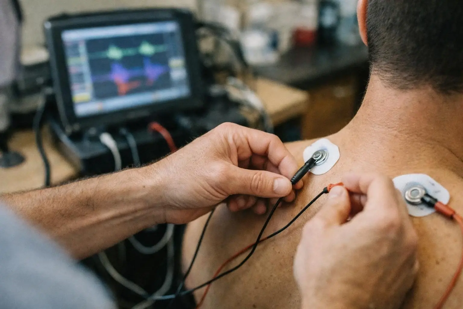
During an EMG, a healthcare provider places electrodes on the skin over a muscle or inserts very thin needles directly into the muscle. These electrodes pick up electrical activity when the muscle is at rest and when it contracts. The signals are displayed on a screen and may also produce sounds. Normal muscles produce predictable patterns of electrical activity. While abnormal patterns can indicate nerve damage, muscle disease. Or other problems.

How Electromyography Works?

An EMG test has two main parts: nerve conduction studies and needle EMG. Nerve conduction studies measure how fast and strong electrical signals travel through a nerve. Small electrodes are placed on the skin over a nerve. And a mild electrical pulse is sent through the nerve. The response is recorded to see if the nerve is functioning properly.

Needle EMG involves inserting a thin needle electrode into a muscle. The needle picks up electrical activity from the muscle fibers. The provider asks the patient to relax or gently contract the muscle while recording the signals. Abnormal electrical patterns, such as reduced activity or unusual spikes, can indicate muscle or nerve damage. The test is usually done in a clinic or hospital and takes about 30 to 60 minutes.

The results of an EMG are interpreted by a specialist, such as a neurologist or chiropractic neurologist. The specialist looks for patterns that suggest conditions like carpal tunnel syndrome, herniated discs, muscle diseases. Or nerve injuries. EMG results are often combined with other tests, like imaging scans, to give a complete picture of a patient’s condition.

Why Electromyography Matters?

How Electromyography applies to Chiropractor services in Brentwood Estates, United States—practical illustration

EMG is a valuable tool because it provides direct information about muscle and nerve function. Unlike X-rays or MRIs, which show the structure of bones and tissues, EMG shows how well nerves and muscles are working. This helps healthcare providers identify the cause of symptoms like pain, tingling. Or weakness. For example, if a patient has numbness in their hand, EMG can determine if the problem is coming from a pinched nerve in the neck, wrist. Or somewhere else.

EMG results guide treatment decisions. If the test shows nerve damage, a chiropractor or doctor may recommend specific therapies, such as spinal adjustments, physical therapy. Or lifestyle changes. Knowing the exact location and severity of a problem helps avoid unnecessary treatments and ensures the right care is provided. EMG is also used to monitor the progress of treatment over time.

When Electromyography Matters Most?

EMG is most useful when patients have symptoms that suggest nerve or muscle problems. Common reasons for an EMG include unexplained muscle weakness, numbness, tingling. Or pain. For example, someone with sciatica—pain that radiates down the leg—may have an EMG to check if a herniated disc is pressing on a nerve. Similarly, a person with carpal tunnel syndrome may have an EMG to confirm nerve compression in the wrist.

The test is also important for patients recovering from injuries or surgeries. EMG can show if nerves are healing properly or if muscles are regaining strength. In some cases, EMG is used for legal or insurance purposes, such as documenting the extent of nerve damage after a car accident. Because EMG provides objective data, it helps ensure patients receive the right diagnosis and treatment for their condition.

How to Evaluate Electromyography?

Related Concepts Compared

Electromyography vs. Nerve Conduction Study (NCS)

NCS measures how fast electrical signals travel through nerves. While EMG records electrical activity in muscles. Both tests are often done together.

Electromyography vs. MRI (Magnetic Resonance Imaging)

MRI shows the structure of bones, muscles. And nerves. While EMG shows how well muscles and nerves are functioning.

Expert Note

EMG is highly sensitive to nerve and muscle dysfunction. But interpretation requires experience. Small changes in electrode placement or muscle contraction can affect results. So it’s important to work with a trained specialist.

Common Mistakes or Myths About Electromyography

  • Thinking EMG is painful—while needle EMG may cause mild discomfort, it is generally well-tolerated.
  • Assuming EMG is the same as an MRI—EMG tests function. While MRI tests structure.
  • Expecting immediate results—EMG data must be interpreted by a specialist, which takes time.
  • Believing EMG can diagnose all types of pain—it is best for nerve or muscle-related issues.

Electromyography in Practice: A Real-World Example

A patient visits a chiropractor with persistent lower back pain and tingling in their left leg. The chiropractor suspects a pinched nerve in the spine. An EMG is performed, which shows reduced electrical activity in the leg muscles and slowed nerve signals. These findings confirm nerve compression. And the chiropractor recommends spinal adjustments and physical therapy to relieve pressure on the nerve.

Related Services

Related Terms

Nerve Compression

Nerve Compression is a condition where a nerve is squeezed or pressed by surrounding tissues like bones, muscles. Or tendons. This pressure can disrupt nerve function, causing pain, tingling, numbness. Or weakness in the affected area. Nerve Compression often occurs in the spine, wrists. Or elbows and may result from injury, repetitive motion.

Chiropractic Care

Chiropractic Care is a health profession focused on diagnosing and treating musculoskeletal disorders, especially those related to the spine. Chiropractic Care uses hands-on spinal adjustments and other manual techniques to improve joint function, reduce pain. And support the body’s natural ability to heal. It's often used for back pain, neck pain, headaches.

Board Certified Chiropractic Neurologist

Board Certified Chiropractic Neurologist is a chiropractor who has completed additional postgraduate training and passed rigorous exams in neurology, earning certification from the American Chiropractic Neurology Board. This certification demonstrates advanced expertise in diagnosing and treating neurological conditions using non-surgical, drug-free methods.

Disc Herniation

Disc Herniation is a spinal condition where the soft inner gel of a spinal disc leaks through a tear in its tough outer layer. This leakage can press on nearby nerves, causing pain, numbness. Or weakness in the back, neck, arms. Or legs. Disc herniations often occur in the lower back or neck due to injury, aging. Or strain.

Range of Motion

Range of Motion is the full movement potential of a joint, typically measured in degrees of a circle. It shows how far a joint can move in different directions, like bending, straightening. Or rotating. Healthy joints have a normal range. While injuries or conditions may limit it. Chiropractors and doctors use this measure to assess joint health and track recovery.

ChiropractorBrentwoodEstates.link

Have Questions About Electromyography?

Contact ChiropractorBrentwoodEstates.link for practical guidance on Electromyography and related chiropractor work in Brentwood Estates.

+1 615-777-0624